


I research small fossil teeth; VERY small fossil teeth. At times these fossil teeth can be submillimeter in size. While a benefit of this is that they do not take up much storage space, one downside is that they can be somewhat difficult to see and study. As I discussed in a previous blog post sometimes it is difficult to even find them in the first place. An obvious solution to the problem of studying these tiny teeth is the microscope. However, there are still problems with viewing teeth of this micro size. Often when looking at these teeth through the microscope, not all of the tooth will be in focus at once due to the narrow focal point. This makes studying these small teeth difficult, as a researcher can only view a small portion of the tooth’s surface at a time. Thankfully, there is a solution to this problem.
By taking a series of photos at different “elevations” along the surface of the tooth, it is possible to create an in-focus image. This is accomplished with a computer program that seamlessly blends or “stacks” these photos together. What you wind up with is an in-focus image that is the result of combining all the focused points of each photo together. In general, the more images you take of the tooth and stack together, the better your final photo. For demonstration purposes please see figures 1-4.
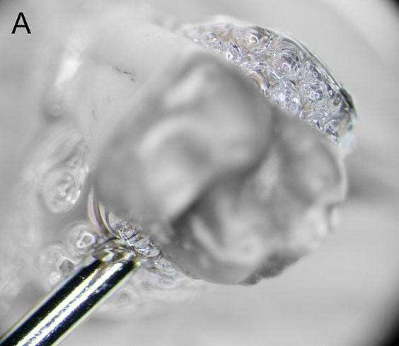
Figure 1: Level “A” is the lowest in-focus slice. Note that only the metal pin (lower left) and the bubbled glue and pin head (upper right) are in focus.
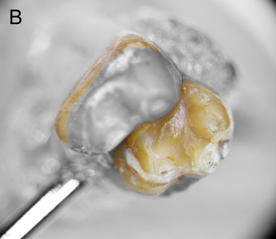
Figure 2: Level “B” is the middle in-focus slice. Note that only the in color sections of the tooth are in focus.
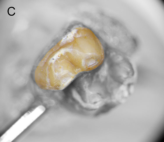
Figure 3: Level “C” is the top in-focus slice. Note that only the in color sections of the tooth are in focus.
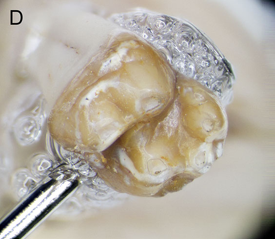
Figure 4: This is the final “stacked” image and is the result of putting all the other images together. Essentially, A + B + C = D. Note that now the entire tooth is in focus.